The aortocoronary bypass is a surgical procedure that is bypassing coronary artery stenosis using autologous arterial or venous grafts. They are harvested from patient body regions where they are not truly necessary. Grafts bypass blood flow from above to under the stenosis, delivering the blood for the underlying myocardium.

Bypass surgery provides the pain-free life in patients with the chest angina, prevents myocardial infarction, and causes further myocardial damage. Due to the high coronary artery disease prevalence, aortocoronary bypass used to be the most performed surgery worldwide.

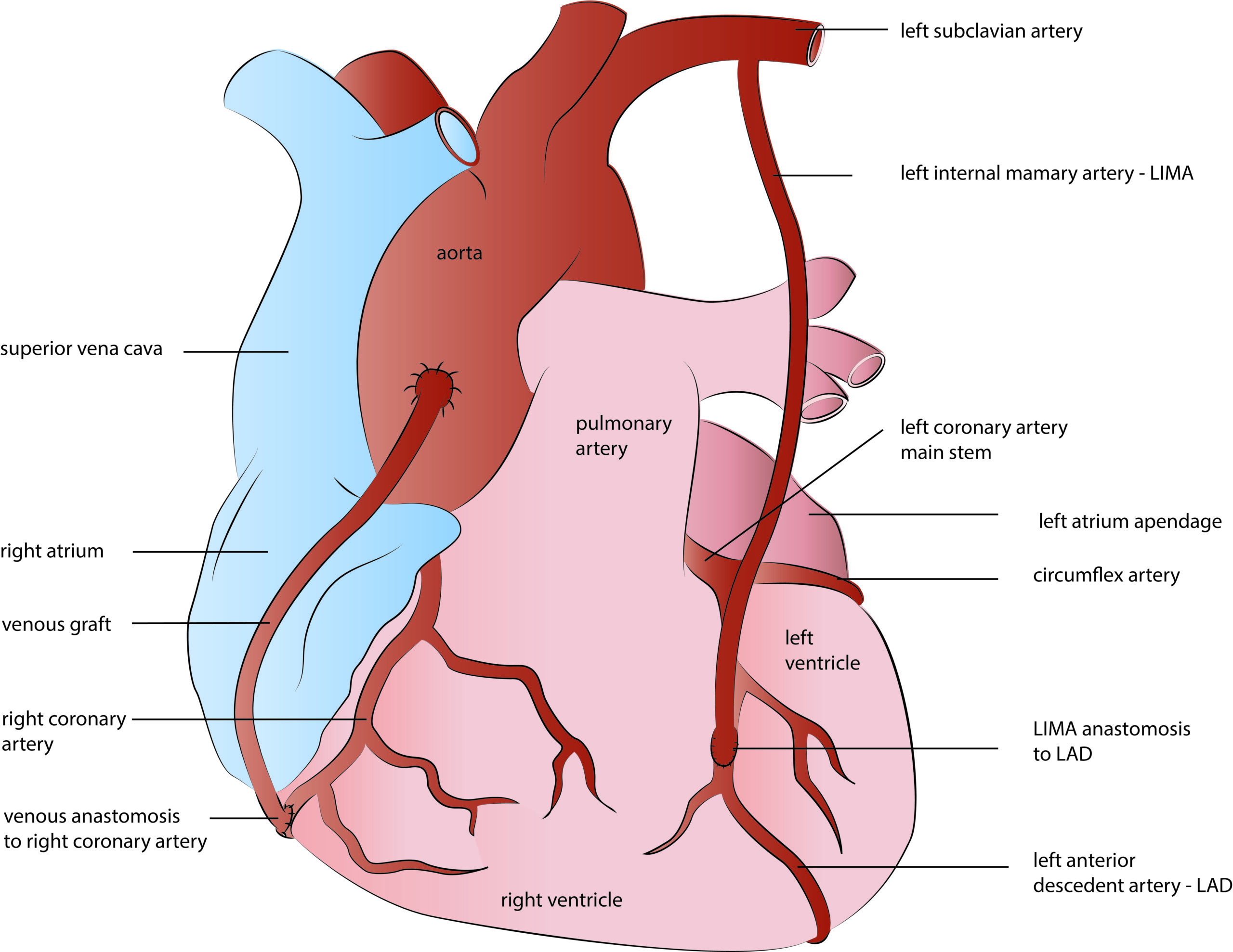
There are two types of blood vessels or grafts used for coronary bypasses: arterial and venous. Venous grafts are harvested from the so-called great saphenous vein, which is a superficial vein from inside your leg. Due to wall anatomy and histology, vein grafts are of inferior quality and, therefore, less durable than arterial grafts. In contrast, arterial grafts are long-lasting and provide superior life quality and long-term survival. Left internal mammary artery – LIMA, is the most commonly used arterial graft. It is harvested from inside of the chest wall and generally used for the left anterior descendent coronary artery bypass. Due to the excellent long-term results, LIMA became a gold standard in coronary surgery today, providing more than 90 % of graft patency rate in the long term follow up studies.

Apart from LIMA, other arteries like right mammary or radial artery- RIMA, are used in coronary surgery today, providing either multiple or total arterial heart revascularization (TAR). The radial artery is a forearm blood vessel frequently used in younger patients as a graft for CABG surgery. Of course, the collateral forearm arteries are previously tested if capable of providing adequate blood supply to hand once the radial artery harvesting.

CORONAROGRAPHY

Coronarography is the imaging technique used to evaluate coronary artery disease anatomy. That is an invasive cath lab procedure performed by tiny catheters. They are introduced into the heart through forearm arteries and used to inject contrast into the coronary artery ostia. Injected contrast fulfills the coronary artery branches and reveals eventual stenosis, location, and severity. That is a gold diagnostic standard for coronary patients, necessary to choose the optimal treatment among the angioplasty, bypass surgery, or medieval therapy.

Before coronarography, we must have clear proof or evidence of chest angina, infarcts, or treadmill test that is positive for coronary artery disease. If not, treadmill or pharmacological stress tests have to be done to support the diagnosis. Patients older than 40 years, patients with significant coronary disease risk factors, and those who have scheduled other than bypass cardiac surgery procedures are indicated for coronarography as well. MSCT exam could be used as a screening test for coronary artery disease in a low-risk patient.